Teena Bansal*, Rajmala Jaiswal**, Manju Bala***
*Assistant Professor; **Professor; ***Senior Resident
Department of Anesthesiology & Critical Care, Pt. B.D. Sharma University of Health Sciences, Rohtak -124001 Haryana, (India)
Correspondence: Dr. Teena Bansal, 2/8 FM, Medical Campus, PGIMS, Rohtak-124001, Haryana, (India); E-mail: aggarwalteenu@rediffmail.com
Citation: Bansal T, Jaiswal R, Bala M. Double upper incisor teeth as a rare cause of difficult intubation. Anaesth Pain & Intensiv Care 2016;20(1):112-113
Key words: Intubation; Laryngeal mask airway; Laryngoscopy
A 32 years old female weighing 50 kg, ASA I was scheduled for cholecystectomy. Her preoperative airway examination revealed bucked teeth, mouth opening 4 cm, mallampati grade II with normal neck mobility and no other obvious airway deformity. General anesthesia was planned for the procedure. In operating room, routine anesthesia was induced with fentanyl 100 µg and thiopentone sodium 250 mg. Mask ventilation was assessed and muscle relaxation was achieved with vecuronium 5 mg. On direct laryngoscopy with Macintosh size 3 blade, we noticed that patient had double upper incisors (Figure). Only epiglottis could be visualized even with external laryngeal manipulate. Two intubation attempts with intubating stylet led to esophageal intubation. Intubating laryngeal mask airway was attempted but failed. Face mask ventilation was still adequate and vitals were stable. Thereafter an attempt was made to insert Proseal laryngeal mask airway (PLMA) No 3. It was appropriately placed in single attempt as judged by adequate chest rise and PetCO2 waveform. Anesthesia was maintained with isoflurane in oxygen and nitrous oxide mixture and maintenance doses of vecuronium. Surgery proceeded uneventfully. At the end of surgery, relaxation was reversed and PLMA was removed. Postoperative course was uneventful.
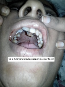
Figure: Double row of upper incisors
Difficult intubations are usually caused by anatomic abnormalities such as micrognathia, limited jaw motion or congenital syndromes. Other causes of difficult intubations include obesity, acromegaly, cervical spine problems and rheumatoid arthritis.1 When anticipated, it allows time and preparation to ensure patient safety. Unanticipated difficult intubation can be challenging at times as in present case. The clinical value of bedside screening methods for predicting difficult intubation remains limited.2-4 Direct laryngoscopic intubation is difficult in 1-4% and impossible in 0.05-0.35% of patients despite an apparently normal pre-operative assessment.3 The unanticipated difficult intubation is probably the most important cause of major anesthesia related morbidity. The ASA difficult airway algorithm recommends using a laryngeal mask airway to secure ventilation and oxygenation (after failed attempts of optimized direct laryngoscopy and change of laryngoscope blades) and also as an intubation conduit with or without fiberoptic guidance in such cases.5 Although various alternative options of intubation (fiberoptic intubation, light wand guided intubation, retrograde intubation, blind nasal intubation) have been described in difficult intubation scenario, we chose LMA, being the most familiar and accessible option, in the given scenario. The other options described require great clinical skills and experience before they can be used in emergency situation.
In our patient there were double upper incisors in same socket, one in front of other, which changed the axis of laryngoscope. This arrangement of teeth could not be noticed during routine preanesthetic checkup with patient in sitting, neutral head position. Perhaps preanesthetic checkups should include checking such conditions as a potential cause of anticipated difficulty in intubation.
REFERENCES
*Assistant Professor; **Professor; ***Senior Resident
Department of Anesthesiology & Critical Care, Pt. B.D. Sharma University of Health Sciences, Rohtak -124001 Haryana, (India)
Correspondence: Dr. Teena Bansal, 2/8 FM, Medical Campus, PGIMS, Rohtak-124001, Haryana, (India); E-mail: aggarwalteenu@rediffmail.com
Citation: Bansal T, Jaiswal R, Bala M. Double upper incisor teeth as a rare cause of difficult intubation. Anaesth Pain & Intensiv Care 2016;20(1):112-113
Key words: Intubation; Laryngeal mask airway; Laryngoscopy
A 32 years old female weighing 50 kg, ASA I was scheduled for cholecystectomy. Her preoperative airway examination revealed bucked teeth, mouth opening 4 cm, mallampati grade II with normal neck mobility and no other obvious airway deformity. General anesthesia was planned for the procedure. In operating room, routine anesthesia was induced with fentanyl 100 µg and thiopentone sodium 250 mg. Mask ventilation was assessed and muscle relaxation was achieved with vecuronium 5 mg. On direct laryngoscopy with Macintosh size 3 blade, we noticed that patient had double upper incisors (Figure). Only epiglottis could be visualized even with external laryngeal manipulate. Two intubation attempts with intubating stylet led to esophageal intubation. Intubating laryngeal mask airway was attempted but failed. Face mask ventilation was still adequate and vitals were stable. Thereafter an attempt was made to insert Proseal laryngeal mask airway (PLMA) No 3. It was appropriately placed in single attempt as judged by adequate chest rise and PetCO2 waveform. Anesthesia was maintained with isoflurane in oxygen and nitrous oxide mixture and maintenance doses of vecuronium. Surgery proceeded uneventfully. At the end of surgery, relaxation was reversed and PLMA was removed. Postoperative course was uneventful.
Figure: Double row of upper incisors
Difficult intubations are usually caused by anatomic abnormalities such as micrognathia, limited jaw motion or congenital syndromes. Other causes of difficult intubations include obesity, acromegaly, cervical spine problems and rheumatoid arthritis.1 When anticipated, it allows time and preparation to ensure patient safety. Unanticipated difficult intubation can be challenging at times as in present case. The clinical value of bedside screening methods for predicting difficult intubation remains limited.2-4 Direct laryngoscopic intubation is difficult in 1-4% and impossible in 0.05-0.35% of patients despite an apparently normal pre-operative assessment.3 The unanticipated difficult intubation is probably the most important cause of major anesthesia related morbidity. The ASA difficult airway algorithm recommends using a laryngeal mask airway to secure ventilation and oxygenation (after failed attempts of optimized direct laryngoscopy and change of laryngoscope blades) and also as an intubation conduit with or without fiberoptic guidance in such cases.5 Although various alternative options of intubation (fiberoptic intubation, light wand guided intubation, retrograde intubation, blind nasal intubation) have been described in difficult intubation scenario, we chose LMA, being the most familiar and accessible option, in the given scenario. The other options described require great clinical skills and experience before they can be used in emergency situation.
In our patient there were double upper incisors in same socket, one in front of other, which changed the axis of laryngoscope. This arrangement of teeth could not be noticed during routine preanesthetic checkup with patient in sitting, neutral head position. Perhaps preanesthetic checkups should include checking such conditions as a potential cause of anticipated difficulty in intubation.
REFERENCES
- Gupta S, Sharma R, Jain D. Airway assessment: predictors of difficult airway. Indian J Anaesth 2005;49:257-62.
- Shiga T, Wajima Z, Inoue T, Sakamoto A. Predicting difficult intubation in apparently normal patients: a meta –analysis of bedside screening test performance. Anesthesiology. 2005 Aug;103(2):429-37. [PubMed] [Free full text]
- Karkouti K, Rose DK, Wigglesworth D, Cohen MM. Predicting difficult intubation: a multivariable analysis. Can J Anesth. 2000 Aug;47(8):730-9. [PubMed]
- Arne J, Descoins P, Fusciardi J, Ingrand P, Ferrier B, Boudigues D, et al. Preoperative assesment for difficult intubation in general and ENT surgery: predictive value of a clinical multivariate risk index. Br J Anaesth. 1998 Feb;80(2):140-146. [PubMed] [Free full text]
- Apfelbaum JL, Hagberg CA, Caplan RA, Blitt CD, Connis RT, Nickinovich DG, et al. Practice guidelines for management of the difficult airway: an updated report by the American Society of Anesthesiologists Task Force on Management of the Difficult Airway. Anesthesiology. 2013 Feb;118(2):251-70. [PubMed] [Free full text]