Subha Teresa Jose Vazhakalayil 1 , Sakshi Sakshi Gupta 2
Authors affiliations:
Citation: Vazhakalayil STJ, Gupta SS. Dual role of the Montgomery Tube as a tracheal stent and function. Anaesth. pain intensive care 2025;29(7):823-824. DOI: 10.35975/apic.v29i7.2981
Received: August 07, 2025; Accepted: October 02, 2025
Dear editor:
We wish to present our findings regarding anesthetic management in the context of Montgomery T-tube insertion in an adult patient exhibiting subglottic stenosis.1 Given the rarity of such cases and the complexities associated with this specialized airway device, we anticipate that this documentation will resonate with anesthesiologists engaged in airway surgical interventions.2
The Montgomery T-tube, initially introduced by William Montgomery in the 1960s, is a silicone, uncuffed T-shaped apparatus employed to bolster the trachea during laryngotracheoplasty.3 It comprises a vertical intraluminal limb and a horizontal extraluminal limb extending through the tracheostomy stoma.4 This device fulfils a dual function: acting as a stent for the airway while also serving as a tracheostomy tube to facilitate respiration and secretion management.5
We describe the case of a 35-year-old female patient who presented with dyspnea and was diagnosed with Grade 3 subglottic stenosis at the C4–C5 vertebral level. Her medical history revealed significant cervical spine trauma due to a vehicular accident, prior anterior cervical discectomy and fusion, prolonged intubation, and a history of tuberculosis lymphadenitis 2 years ago.
Subsequent to an emergency tracheostomy and medical optimization, the patient was scheduled for definitive airway reconstruction with the insertion of a Montgomery T-tube distal to the stenotic segment to ensure a patent airway and avoid bypass obstruction. Standard ASA monitors were applied. Anesthesia was initiated intravenously and sustained with a combination of oxygen, nitrous oxide, sevoflurane, vecuronium, and a continuous dexmedetomidine infusion. The tracheostomy tube was replaced with a 7.5 mm flexometallic cuffed tube to ensure controlled ventilation.
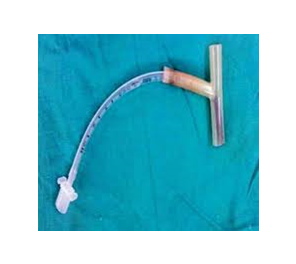
Figure 1: ETT connected to the outer limb of the T-tube for ventilation
To facilitate T-tube insertion, the surgeon utilized a 14 Fr Foley catheter, which was introduced via direct laryngoscopy to occlude the upper limb of the T-tube. The breathing circuit was subsequently connected to the extratracheal limb with the use of a 6.5 mm endotracheal tube connector. This technique effectively ensured adequate ventilation throughout the apneic phase of the procedure. Neuromuscular blockade was successfully reversed, and the patient was transferred to the Surgical Intensive Care Unit (SICU) following postoperative recovery. The unique configuration of the Montgomery T-tube poses several challenges for the anesthesiologists:
Conflict of interest
Nil declared by the authors.
Authors contribution
Both authors took part in the management of the case and drafting this manuscript
Authors affiliations:
- Subha Teresa Jose Vazhakalayil, Dr. D. Y. Patil Medical College Hospital and Research Centre Sant, Tukaram Nagar Pimpri, Pune, India; Email: dr.subhajose@gmail.com
- Sakshi Gupta, Dr. D. Y. Patil Medical College, Tukaram Nagar Pimpri, Pune, India; Email: sakshig536@gmail.com; {ORCID:0009-0007-89385689}
Citation: Vazhakalayil STJ, Gupta SS. Dual role of the Montgomery Tube as a tracheal stent and function. Anaesth. pain intensive care 2025;29(7):823-824. DOI: 10.35975/apic.v29i7.2981
Received: August 07, 2025; Accepted: October 02, 2025
Dear editor:
We wish to present our findings regarding anesthetic management in the context of Montgomery T-tube insertion in an adult patient exhibiting subglottic stenosis.1 Given the rarity of such cases and the complexities associated with this specialized airway device, we anticipate that this documentation will resonate with anesthesiologists engaged in airway surgical interventions.2
The Montgomery T-tube, initially introduced by William Montgomery in the 1960s, is a silicone, uncuffed T-shaped apparatus employed to bolster the trachea during laryngotracheoplasty.3 It comprises a vertical intraluminal limb and a horizontal extraluminal limb extending through the tracheostomy stoma.4 This device fulfils a dual function: acting as a stent for the airway while also serving as a tracheostomy tube to facilitate respiration and secretion management.5
We describe the case of a 35-year-old female patient who presented with dyspnea and was diagnosed with Grade 3 subglottic stenosis at the C4–C5 vertebral level. Her medical history revealed significant cervical spine trauma due to a vehicular accident, prior anterior cervical discectomy and fusion, prolonged intubation, and a history of tuberculosis lymphadenitis 2 years ago.
Subsequent to an emergency tracheostomy and medical optimization, the patient was scheduled for definitive airway reconstruction with the insertion of a Montgomery T-tube distal to the stenotic segment to ensure a patent airway and avoid bypass obstruction. Standard ASA monitors were applied. Anesthesia was initiated intravenously and sustained with a combination of oxygen, nitrous oxide, sevoflurane, vecuronium, and a continuous dexmedetomidine infusion. The tracheostomy tube was replaced with a 7.5 mm flexometallic cuffed tube to ensure controlled ventilation.
Figure 1: ETT connected to the outer limb of the T-tube for ventilation
To facilitate T-tube insertion, the surgeon utilized a 14 Fr Foley catheter, which was introduced via direct laryngoscopy to occlude the upper limb of the T-tube. The breathing circuit was subsequently connected to the extratracheal limb with the use of a 6.5 mm endotracheal tube connector. This technique effectively ensured adequate ventilation throughout the apneic phase of the procedure. Neuromuscular blockade was successfully reversed, and the patient was transferred to the Surgical Intensive Care Unit (SICU) following postoperative recovery. The unique configuration of the Montgomery T-tube poses several challenges for the anesthesiologists:
- The airway is partially shared with the surgical team, leading to a potential loss of airway control during insertion.
- The extratracheal limb lacks a conventional 15 mm connector, necessitating modifications.
- Considerable loss of anesthetic gases may occur through the open proximal limb of the intratracheal segment.
Conflict of interest
Nil declared by the authors.
Authors contribution
Both authors took part in the management of the case and drafting this manuscript
REFERENCES
- Montgomery WW. T-tube tracheal stent. Arch Otolaryngol. 1965;82(3):320-1. DOI: 10.1001/archotol.1965.00760010322023
- Das A, Chawla N, Sharma B, Samujh R, Kumar P. Anaesthetic challenges in a child with a Montgomery T-tube in situ. Indian J Anaesth. 2012;56(3):296-8. DOI:10.4103/ija.IJA_293_20
- Guha A, Mostafa SM, Kendall JB. The Montgomery T-tube: anaesthetic problems and solutions. Br J Anaesth. 2001 Nov;87(5):787-90. DOI: 10.1093/bja/87.5.787
- Massard G, Rouge C, Dabbagh A, Kessler R, Hentz JG, Roeslin N, et al. Tracheobronchial stenting for postoperative tracheal stenosis. Ann Thorac Surg. 1996;62(6):1665-9. DOI: 10.1016/0003-4975(96)00083-5
- Greer JR, Kwo J, Macario A, Brock-Utne JG. Anesthesia for tracheal resection and reconstruction: the two-lung ventilation technique. Can J Anaesth. 1999;46(6):588-91.
- Phillips PS, Kubba H, Hartley BE, Albert DM. The use of the Montgomery T-tube in difficult paediatric airways. Int J Pediatr Otorhinolaryngol. 2006 Jan;70(1):39-44. DOI: 10.1016/j.ijporl.2005.05.008